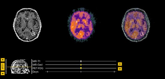
A 73 year old right-handed man sought
medical attention because of a grand mal seizure and progressive difficulty
with speech. Brain biopsy in 3/95 revealed grade II astrocytoma; however,
because of his age and speed of progression, the pathological result was
thought to be artifactually benign and due to sampling error. He underwent
conventional whole brain radiation consisting of 6440 cGys given in 35
treatments. He tolerated his radiation therapy well, but attempts to wean his
steroids resulted in prompt seizure recurrence. Concern of tumor recurrence vs.
radiation necrosis prompted evaluation with PET.

The T1-weighted precontrast MR(not
shown) demonstrated an area of low signal intensity throughout the left
parietal-occipital area which was new. A previous study had shown only
postoperative changes. Following the administration of contrast material(gadolinium-DTPA), signal intensity was much enhanced, as seen in image 59 and
surronding slices, suggesting either tumor recurrence or radiation necrosis.
This finding also represented a change when compared to the previous contrast
enhanced MR. The PET scan demonstrated high activity in this region, suggestive
of tumor recurrence, and effectively ruling out radiation necrosis.
Treatment options included surgical
resection of the area of increased metabolic activity consistent with tumor
recurrence or stereotactic radiosurgery to that same area, either as a primary
treatment or for palliation, if follow-up MR again revealed contrast
enhancement.
Like Thallium SPECT, the PET scan has
been very useful in differentiating tumor recurrence and radiation necrosis. It
also allows specific targeting of the area of recurrence, which may be
surrounded by radiation necrosis in those patients who have received radiation.
Case contributed by Drs. C. D. Sturm
and R. Bucholz, St. Louis University.

