A 51
year old woman sought medical attention because of gradually increasing right
hemiparesis (weakness) and hemianopia(visual loss). At craniotomy(8/90),
left parietal anaplastic astrocytoma was found. A right frontal lesion was
biopsied in 8/94. Recurrent tumor was suspected on the basis of the imaging,
and was confirmed pathologically. Total images available: 1,045. All have
been brought into registration using “Superpose”.
Superpose
is a set of software tools for analysis of medical images. Its major purpose
is to permit the precise comparison of image datasets from different sources
and from different time points. Images from a variety of modalities,
including MR, CT, SPECT, and PET, can be precisely compared with each other.
With Superpose, volume datasets are processed automatically to yield images
which are corrected for differences in pixel size and patient orientation.
This allows, for example, precise comparison of brain functional images with
underlying structure. It also permits accurate comparison of datasets
gathered in a time sequence.
The
superpositioning procedure is entirely retrospective. It does not require
fiducial markers or special patient positioning. The processing of two
typical datasets requires, on average, less than five minutes to complete.
The
superposition algorithm operates on surfaces derived from multi-slice or
volume datasets.
Here
is an example of the pair of surfaces. A surface is derived from a set of
contours which outlines the edge of a volume on a series of planar images.
Contours are generated by an edge detection algorithm based on a user chosen
pixel threshold. This threshold is varied under mouse control until a
suitable edge is determined. Contours in all subsequent slices are
automatically calculated based on parameters of the first. The corresponding
surface consists of a set of triangles spanning neighboring pairs of
contours.
Two
surfaces are superposed by finding the rigid body transformation carrying one
surface into the other which minimizes the volume between them. The
transformation is specified by six parameters: three translational and three
orientational. The transformation which minimizes the volume contained
between the two surfaces is taken to be the transformation relating the two
underlying image volumes.

The
triangulated MR brain surface is drawn in yellow. The red or green vectors represent the displacement
between the two surfaces.
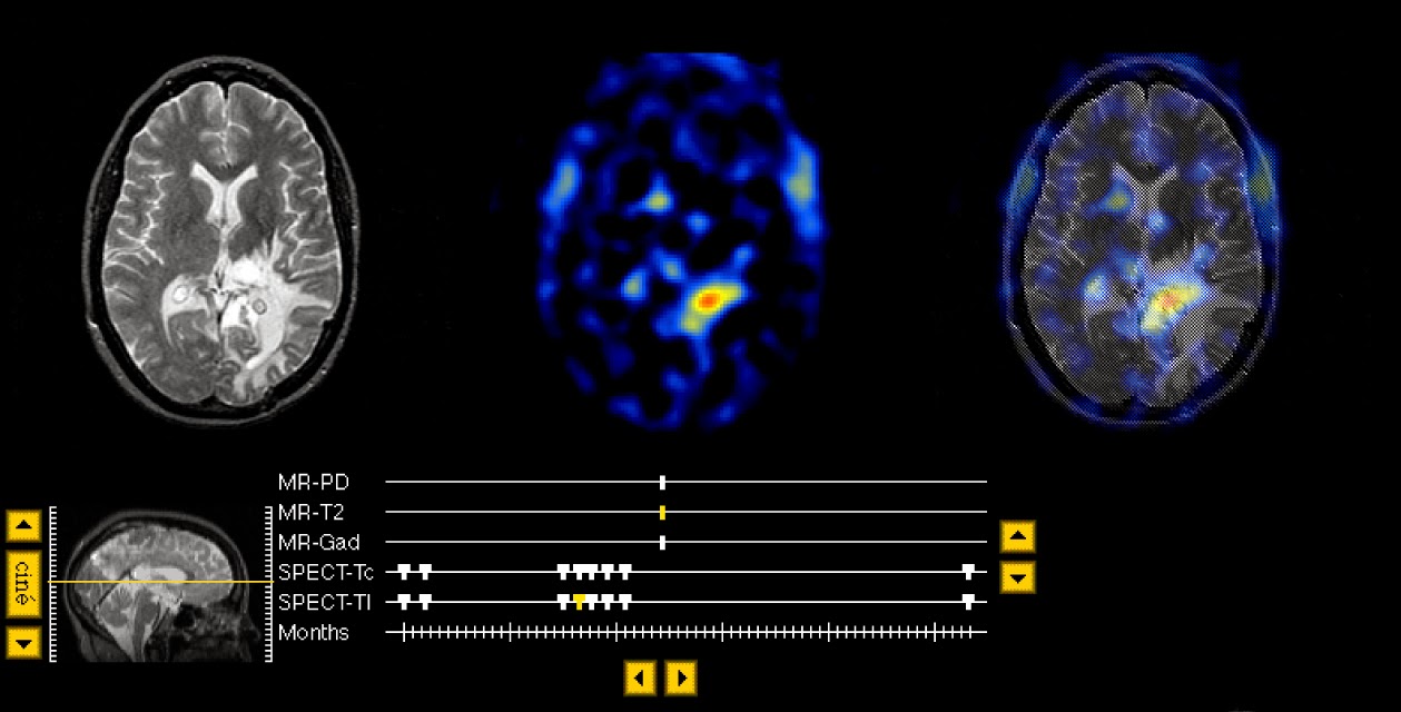
Note particularly:
·
the
evolution of high tumor Thallium uptake, indicating astrocytoma recurrence.
This can best be appreciated by choosing the Thallium study (with the thick
tickmark for overlay) and then clicking the "time" button.
·
the
large region of mixed signal on T2- and PD- weighted MR, only a subset of
which actually corresponds to active tumor.
Tours: Tour 1. Parietal lesion
Another
method of imaging which reflects breakdown of the blood-brain barrier is
shown here. This is an image of cerebral uptake of Thallium-201, a potassium
analog which "leaks" into the brain at sites of active tumor
growth. Note the 2 large foci of red-colored activity in the sites of 201-Tl
uptake. Compare this with the previous tour stop to see the correspondence of
Thallium-201 images with gadolinium MR. These images are "in
register", and are therefore samples of the same slice of brain. On the
next stop, we will look at a higher slice of the same stack, to see the more
superior extent of the active portion of the lesion.
 Tour 2. Edema
This
tour will examine the cerebral edema which corresponds generally to the high
signal extending from the center of the mass through surrounding white
matter. On this proton-density-weighted image, the high signal corresponding
to edema respects the gray-white junction, but tends to spare the
"u"-fibers. This is a common observation in the white matter
reaction to malignant neoplasia, so-called "vasogenic" edema.
Choose a spatial movie (by clicking "cine" next to the sagittal
image) of the proton density stack to see the extent of the edema.
 Tour 3. Contrast Enhancement
The hypopheseal or pituitary stalk is bright because it lacks the barrier found throughout most of the brain. This structure, also known as the infundibulum, is often grouped with other "circumventricular organs" which share the property of being outside the blood-brain-barrier.
|


