Longitudinal magnetic resonance study: a year in the life
of a patient with the chronic progressive form of the disease. These data were
assembled using Superpose, a method of image registration.
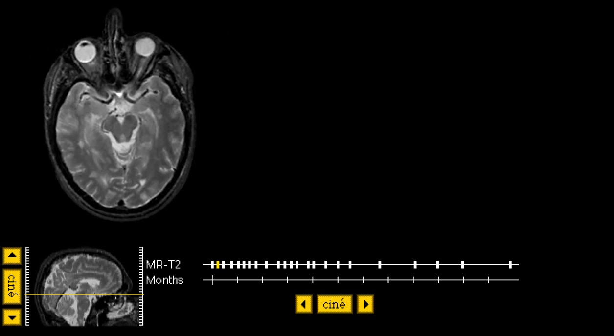
This is a tour of the lesions of multiple sclerosis. Look at the "temporal" movie (time-cine) to see any changes occurring during one year's time. Look at the "spatial" movie (choose "cine", next to the sagittal image) to see the whole brain. There are a few small regions of high signal in temporal white matter, but, at this level, the image is mostly normal. On our next stop, we will see a larger lesion.
This is a tour of the lesions of multiple sclerosis. Look at the "temporal" movie (time-cine) to see any changes occurring during one year's time. Look at the "spatial" movie (choose "cine", next to the sagittal image) to see the whole brain. There are a few small regions of high signal in temporal white matter, but, at this level, the image is mostly normal. On our next stop, we will see a larger lesion.
.jpg)
Here are a few sample "time-lapse" images(MPEGs) showing MS lesion evolution over one year's time.
supraventricular
supra-thalamic
thalamus
midbrain
Look
at the large round white spot in the right frontal region. This is a relatively
new lesion, and you can see how it enlarges very rapidly over the next weeks.
Look at the timeline cine. With time, the lesion enlarges, there is a
"halo" of white (high) signal which surrounds the lesion. This
probably represents the edema which forms in reaction to the acute damage. At
the end of the movie, you can see that the lesion has nearly disappeared, with
another lesions appearing.

Tour2
Here are a few small lesions in the lower pons. On
this tour, we will review the morphology and location of smaller lesions.


The superpositioning procedure is entirely retrospective. It does not require fiducial markers or special patient positioning. The processing of two typical datasets requires, on average, less than five minutes to complete.
The superposition algorithm operates on surfaces derived from multi-slice or volume datasets.
Here is an example of the pair of surfaces. A surface is derived from a set of contours which outlines the edge of a volume on a series of planar images. Contours are generated by an edge detection algorithm based on a user chosen pixel threshold. This threshold is varied under mouse control until a suitable edge is determined. Contours in all subsequent slices are automatically calculated based on parameters of the first. The corresponding surface consists of a set of triangles spanning neighboring pairs of contours.
Two surfaces are superposed by finding the rigid body transformation carrying one surface into the other which minimizes the volume between them. The transformation is specified by six parameters: three translational and three orientational. The transformation which minimizes the volume contained between the two surfaces is taken to be the transformation relating the two underlying image volumes.

